


Choosing a Diagnostic Medical Sonography Program means preparing for a field that combines science, technology, patient care, and hands-on imaging. Students study the human body, learn how sound waves create diagnostic images, practise scanning techniques, and develop clinical skills for patient care.
At Pharma-Medical Science College of Canada, sonography education combines classroom learning, laboratory training, and clinical experience. Understanding the subjects can help you see how the curriculum supports work in diagnostic imaging.
What Subjects Are Included in a Diagnostic Medical Sonography Program?
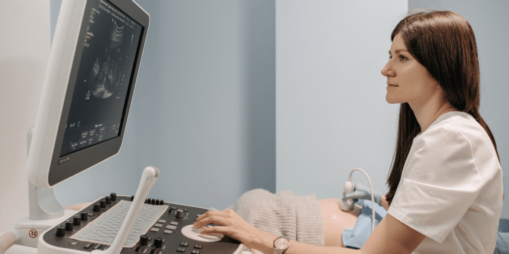
A Diagnostic Medical Sonography Program typically includes anatomy and physiology, pathophysiology, medical terminology, ultrasound physics, instrumentation, specialty imaging, patient care, scanning laboratories, and clinical training. Together, these subjects help students understand human anatomy, produce diagnostic images, and develop practical patient care skills.
The Diagnostic Medical Sonography diploma program at Pharma-Medical includes theoretical and clinical training across several sonography areas, including abdomen and small parts, gynecology, obstetrics, abdominal vascular imaging, minor musculoskeletal sonography, and pediatric sonography.
Key Takeaway
Think of sonography education in four connected layers: science explains the body, ultrasound physics explains the image, specialty courses explain what to examine, and clinical training teaches you how to apply those skills with patients.
A useful way to think about the diagnostic medical sonography curriculum is this: science explains the body, ultrasound physics explains the image, specialty courses explain what to examine, and clinical training teaches you how to bring all of that together.
Foundational science subjects build your imaging knowledge
Medical sonography education begins with the academic foundation needed to understand anatomy, disease, and clinical language. These subjects become increasingly practical once students begin scanning.
Anatomy and physiology
Anatomy teaches the location, structure, and relationships of organs, tissues, vessels, and body systems. Physiology focuses on how those structures normally function.
For a sonography student, this knowledge is highly visual. When scanning the abdomen, you need to know where organs and major vessels should appear in relation to one another. Normal anatomy becomes the reference point for recognizing changes that may require closer assessment.
Pathophysiology
Pathophysiology examines how disease or injury changes normal body function. This helps students connect what they know about healthy anatomy with the kinds of changes that may appear during diagnostic imaging.
Sonographers are trained to acquire and document diagnostic images within their scope of practice, not replace the physician responsible for diagnosis. Pathophysiology helps students understand why certain views, measurements, or observations may be clinically important.
Medical terminology and quantitative foundations
Medical terminology helps students understand chart notes, requisitions, anatomical directions, and clinical communication. Mathematics and general physics may also support later work with ultrasound physics, measurements, Doppler calculations, and image optimization.
These foundational subjects are part of the reason diagnostic medical sonography courses can feel academically demanding. Students are learning a healthcare language while also learning technical imaging concepts.
Ultrasound physics and instrumentation explain how the image is created
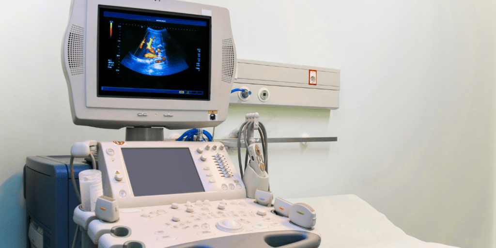
Ultrasound physics is one of the most technical parts of sonography education. Students learn how high-frequency sound travels through tissue, how echoes return to the transducer, and how the ultrasound system converts that information into an image.
Topics can include frequency, wavelength, reflection, attenuation, resolution, transducer selection, Doppler principles, and system controls. Instrumentation lessons connect those principles to the ultrasound machine.
A technically poor image can hide useful information. Sonographers need to understand which controls affect depth, gain, focus, frequency, and other image characteristics.
For students researching ultrasound technology courses, this is an important point to compare. Good training should connect physics to image quality and scanning decisions, rather than treating physics as a subject that exists only for an exam.
Clinical imaging subjects teach you what and how to scan

Specialty imaging courses take foundational science and apply it to specific body areas and clinical questions. This is where students begin to see how different examinations require different scanning approaches.
| Subject Area | What Students Study | Why It Matters |
|---|---|---|
| Anatomy and Physiology | Body structures, organs and normal function | Helps students recognize normal anatomy during scanning |
| Pathophysiology | How disease or injury affects the body | Connects clinical conditions with imaging observations |
| Ultrasound Physics | Sound waves, Doppler principles and image formation | Supports image quality and equipment operation |
| Specialty Imaging | Abdominal, obstetric, gynecologic, vascular and other imaging | Builds examination-specific scanning knowledge |
| Patient Care | Communication, privacy, positioning and safety | Supports professional patient interactions |
| Scanning Labs | Probe handling, controls and image acquisition | Turns classroom knowledge into practical technique |
| Clinical Practicum | Supervised experience in clinical settings | Brings technical and patient-care skills together |
Abdominal and small-parts sonography
Abdominal sonography can involve imaging organs such as the liver, gallbladder, kidneys, pancreas, and related structures. Small-parts sonography may include superficial structures that require careful transducer positioning and image optimization.
Students learn standard scanning planes, anatomical landmarks, measurement techniques, and how patient position can affect image acquisition.
Obstetrics and gynecology
Obstetric and gynecologic sonography focuses on the female pelvis and pregnancy-related imaging. Students may learn pelvic anatomy, fetal development, fetal measurements, and scanning approaches used across different stages of pregnancy.
This area also highlights patient care. Examinations can involve sensitive situations, discomfort, or anxiety, so professionalism, privacy, and communication matter.
Abdominal Vascular and Doppler Imaging
Abdominal vascular sonography uses ultrasound and Doppler techniques to examine blood ships, and assess blood flow in the abdomen. Students learn how vascular anatomy, Doppler principles, and scanning techniques contribute to producing useful diagnostic images.
This subject shows how the curriculum connects: anatomy identifies the vessel, physics explains the Doppler signal, and scanning practice teaches students how to obtain a useful waveform.
Pediatric and minor musculoskeletal sonography
The current Pharma-Medical program also identifies pediatric and minor musculoskeletal sonography among its training areas, exposing students to additional scanning situations and patient needs.
Students interested in a broader diagnostic medical sonography career can review the career guide for sonography in Ontario to see how education connects with potential work settings and professional requirements.
Patient care, communication, ethics, and safety are part of the curriculum
A diagnostic ultrasound appointment is a patient interaction, not simply an imaging task. That is why patient management and communication belong alongside technical subjects.
Students need to explain examinations appropriately, protect privacy, assist with positioning, and respond calmly when a patient is uncomfortable. Infection prevention, workplace safety, documentation, and ethical responsibilities may also be part of clinical preparation.
These skills are easy to underestimate when comparing diagnostic medical sonography programs in Ontario, yet they affect nearly every clinical encounter.
Scanning labs turn theory into repeatable technique
Scanning laboratories give students supervised time with ultrasound equipment before and alongside clinical placement. This is where probe handling, hand-eye coordination, body mechanics, image orientation, and machine controls begin to feel less abstract.
At first, students may focus on finding a structure and keeping the image steady. With practice, attention shifts to image quality, measurements, scanning sequence, and documentation.
This progression is one of the most important parts of medical sonography education. Knowledge needs repetition before it becomes a reliable clinical skill.
A practical point that students sometimes miss: scanning is physical work. Learning good ergonomics early can matter for shoulder, wrist, neck, and back comfort during repeated examinations.
Clinical practicum shows how the subjects work together
Clinical placement is where classroom knowledge, lab practice, communication, and professional behaviour meet. Pharma-Medical currently lists a 32-week clinical practicum in approved clinical settings as part of its program.
During supervised clinical experience, students can apply scanning protocols, work with patients, observe workflow, receive feedback, and build confidence under real clinical conditions. The deeper value is learning how to adapt when patients, time limits, and clinical questions vary.
Students exploring what the field looks like after school may also find the college’s guide to a diagnostic medical sonography career in Canada useful.
Considering a Career in Diagnostic Medical Sonography?
Ask our admissions team about program subjects, admission requirements and upcoming intakes.
Contact UsHow do these subjects prepare you for a sonography career?
The subjects work as a system. Anatomy without scanning practice stays theoretical. Scanning without physics can weaken image optimization, while technical skills without patient communication can make clinical work difficult.
A well-structured program therefore moves students from knowledge to application, with several subjects coming together during a single examination.
Some learners also become interested in related specialties after studying general sonography. Cardiac sonography, for example, focuses specifically on imaging the heart and is commonly offered as a separate training pathway. Students curious about that direction can read about diagnostic cardiac sonography careers.
What should you compare before choosing a sonography program?

Course names alone do not show whether a program is a good fit. Compare how subjects are taught and how students move from theory into scanning practice. A useful review checklist includes program accreditation, admission requirements, laboratory access, clinical practicum structure, areas of sonography covered, instructor experience, and the certification pathways the curriculum is intended to support.
You can also review Pharma-Medical’s article on joining an accredited diagnostic medical sonography program in Toronto for another perspective on what to consider. Review the current sonography course requirements before applying, including academic prerequisites and any health, safety, or documentation requirements for clinical placement.
Start by understanding what you will actually learn
A Diagnostic Medical Sonography Program is built from several connected subjects, including anatomy and physiology, pathophysiology, ultrasound physics, instrumentation, specialty imaging, patient care, scanning labs, and clinical practice. Each subject develops a different part of the sonographer’s skill set.
If you are considering this career path, review the current program structure, admission details, clinical training, and areas of study before making a decision. To ask about Pharma-Medical’s program, upcoming intakes, or the application process, contact the college and speak with the admissions team.
Frequently Asked Questions About Diagnostic Medical Sonography
What subjects are taught in a Diagnostic Medical Sonography Program?
Common subjects include anatomy and physiology, pathophysiology, ultrasound physics and instrumentation, specialty imaging, patient care, scanning laboratories and clinical practicum training.
Is ultrasound physics an important part of sonography training?
Yes. Ultrasound physics helps students understand how sound waves produce images, how Doppler works and how machine settings affect image quality.
Does the Pharma-Medical sonography program include hands-on training?
Yes. The program combines classroom instruction, laboratory training and clinical experience, including a 32-week clinical practicum in approved clinical settings.
How long is the Diagnostic Medical Sonography Program at Pharma-Medical?
Pharma-Medical currently lists the Diagnostic Medical Sonography program as 100 weeks of instructor-led, full-time training and clinical practice, including a 32-week clinical practicum.
What areas of sonography are covered at Pharma-Medical?
The current program covers abdomen and small parts, gynecology, obstetrics, abdominal vascular imaging, minor musculoskeletal sonography and pediatric sonography.
What should I compare when choosing a sonography program in Ontario?
Compare accreditation, admission requirements, curriculum content, laboratory access, clinical practicum opportunities and the professional certification pathways the program is designed to support.